
Constantly expanding the field of perception into dimensions initially hidden from the naked eye, drives science forward. Today, increasingly powerful microscopes let us see into the cells and tissues of living organisms, into the world of microorganisms as well as into inanimate nature. But even the best microscopes have their limits.
“To be able to observe structures and processes down to the nanoscale level and below, we need new methods and technologies,” said Dr. Silvio Fuchs from the Institute of Optics and Quantum Electronics at the University of Jena in Germany. This applies in particular to technological areas such as materials research or data processing.
The new method, which they call Coherence Tomography with Extreme Ultraviolet Light (XCT), emanates from optical coherence tomography (OCT), which has been established in ophthalmology for a number of years, said doctoral candidate Felix Wiesner, the lead author of the study. “These devices have been developed to examine the retina of the eye non-invasively, layer by layer, to create 3-dimensional images.”

At the ophthalmologist, OCT uses infrared light to illuminate the retina. The radiation is selected in such a way the tissue to be examined does not absorb it too strongly and it can end up reflected by the inner structures. However, the physicists at Jena use extremely short-wave UV light instead of long-wave infrared light for their OCT. “This is due to the size of the structures we want to image,” Wiesner said. In order to look into semiconductor materials with structure sizes of only a few nanometers, they only need light with a wavelength of only a few nanometers.
Generating such extremely short-wave UV light (XUV) used to be a challenge and was almost only possible in large-scale research facilities.
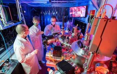
Prof. Dr. Gerhard Paulus, PhD student Felix Wiesner and Dr Silvio Fuchs (from left) in a laser lab of the Institute of Optics and Quantum Electronics at the University of Jena in Germany.
Source: Jens Meyer (University of Jena)
Jena physicists, however, generate broadband XUV in an ordinary laboratory and use what are called high harmonics for this purpose. This is radiation produced by the interaction of laser light with a medium and it has a frequency many times that of the original light. The higher the harmonic order, the shorter the resulting wavelength.
“In this way, we generate light with a wavelength of between 10 and 80 nanometers using infrared lasers,” said Prof. Gerhard Paulus, professor of nonlinear optics at the University of Jena. “Like the irradiated laser light, the resulting broadband XUV light is also coherent, which means that it has laser-like properties.”
In the work described in their current paper, the physicists exposed nanoscopic layer structures in silicon to the coherent XUV radiation and analyzed the reflected light. The silicon samples contained thin layers of other metals, such as titanium or silver, at different depths. Because these materials have different reflective properties from the silicon, they can end up detected in the reflected radiation.
The method is so precise that not only can the deep structure of the tiny samples be displayed with nanometer accuracy, but – due to the differing reflective behavior – the chemical composition of the samples can also end up determined precisely and, above all, in a non-destructive manner.
“This makes coherence tomography an interesting application for inspecting semiconductors, solar cells or multilayer optical components,” Paulus said, adding it could also end up used for quality control in the manufacturing process of such nanomaterials, to detect internal defects or chemical impurities.